Over the past 13 years, a team of individuals from the Fanconi Anemia German Support Group, plus FA researchers and clinicians from the University of Düsseldorf, have conducted a study to determine if oral pre-malignant and malignant lesions can be accurately identified using brush biopsies. These biopsies provide a painless and non-invasive method to obtain oral tissue samples. Researchers collected data from more than 700 participants worldwide. They summarized their results in an article entitled Diagnostic accuracy of brush biopsy-based cytology for early detection of oral cancer and precursors in Fanconi anemia, published in the journal Cancer Cytopathology, February 2020.
Individuals with Fanconi anemia (FA) are at high risk of developing oral cancer. Unlike cancers found in the general population, these FA cancers cannot be treated with chemotherapy because of the underlying DNA repair problem. Therefore, early detection and surgical intervention are essential.
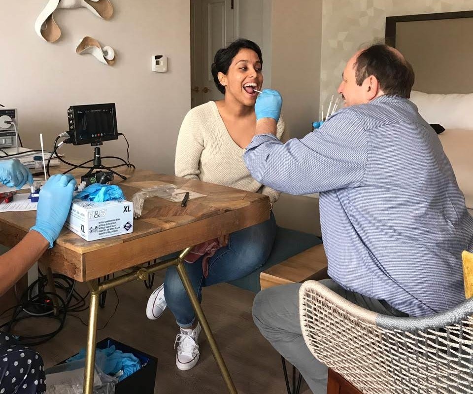
Ralf Dietrich (FA German Family Support Group) takes a sample from Alejandra at the FA Adult Meeting
Oral cancer begins as a precancerous lesion. If a lesion does not heal in four weeks, FA individuals are advised to consult with an otolaryngologist (ENT physician). If the lesion appears suspicious, the physician often performs a biopsy. Could precancerous lesions be detected much earlier, before they become cancerous and without enduring a painful, invasive biopsy?
Dr. Schramm, head of the department of Cytopathology from the University of Düsseldorf, noted that routine use of brush biopsies in the general population is controversial. Dr. Schramm noted: “Especially in a patient cohort like individuals with FA, where changes of the oral mucosa develop frequently, we had to show that this method is accurate.”
The published results show that this method is, indeed, accurate. Using two different methods of analysis, the “sensitivity” of these studies was 100% (ability to correctly identify those with pre-malignant or malignant lesions) and the “specificity” (ability to identify those without disease) was 92.2%. Equally significant, approximately 63% of squamous cell carcinoma and precursor lesions were detected at a non-invasive or early stage, making surgical removal effective.
This is how brush biopsies work: the lesion is swabbed, and if the brush test is negative, the lesion can be monitored clinically, and an invasive biopsy is not necessary. If the test result is ambiguous or positive, the results need further investigation. Dr. Schramm notes: “We don’t want to avoid invasive biopsies completely. But we hope that with the brush biopsy as a screening tool, we will identify those lesions that should be treated by experts.” Dr. Eunike Velleuer, University of Düsseldorf, hopes that this procedure will improve the basic care for individuals with FA who develop visible oral lesions. These lesions are more frequently malignant in FA than in the general population, but only a minority of all visible oral lesions in people with FA is worrisome, which is why it is unnecessary to biopsy all FA oral lesions.
Ralf Dietrich of the German Support Group believes that in the past, many people who developed oral cancer experienced poor survival because they were diagnosed too late. Dietrich is convinced that with routine use of brush biopsies, oral cancer will be diagnosed much earlier and will therefore be more treatable.
Dr. Velleuer sums it up: “Now that we know this methodology is an accurate way to detect early cancers in FA patients, we can look to the future with more hope. We need to make this technology widely accessible so that all individuals with FA can have these screenings performed by their doctors, and learn why people with positive results developed cancer. If we can identify common risk factors and learn more about the nature of those cancers, we can work on prevention.”
Are you interested in participating in the oral screenings study? Team members are available at the FA Family Meeting and Meeting for Adults with FA every year, in addition to numerous gatherings of FA families around the world. The team also visits individuals in their homes to perform these screenings. Please contact Suzanne Planck, FARF Family Services Coordinator, if you’re interested in learning more: Suzanne@fanconi.org, 541-687-4658